Introduction:
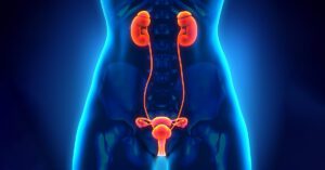
Uretero-pelvic junction (UPJ) obstruction represents a set of obstructive processes that result from multiple etiologic factors. These include:
- Aberrant renal vessel or fibrous band compressing the UPJ.
- Intrinsic narrowing of the UPJ.
- High insertion of the ureter into the renal pelvis.
- Other causes such as stones, previous surgery, infections, iatrogenic injury, etc. may cause secondary UPJ obstruction.
Such an abnormality at the UPJ may hinder the appropriate propagation of peristaltic wave of urine.
Treatment Options:
Historically, open surgical management (pyeloplasty) was the treatment of choice for UPJ obstruction. However, with the introduction of endourology, the treatment options have expanded to include:
- Antegrade percutaneous endopyelotomy
- Retrograde ureteroscopic endopyelotomy
- Balloon with a cutting electrode (Acusise)
Retrograde Ureteroscopic Endopyelotomy
Our first patient underwent this procedure on November 20, 1989. The procedure has been developed on sound endourologic principles of:
- Appropriate instrumentation
- Safe electrocautery use
- Judicious stenting of the UPJ
- Anatomic considerations at the UPJ.
- Urinary tract dynamics
Instrumentation Required for Ureteroscopic Endopyelotomy:
- 11.5 Fr. Uretero- resectoscope (Storz Wolf) with both cold and electrocautery attachments. Not a regular ureteroscope.
- Super-stiff guidewire (Meditech)
- 5 Fr. open-ended catheter.
- Single action pump system (Microvasive)
- Balloon dilator (Olbert preferred)
- Retromax endopyelotomy stent (Microvasive)
- Routine cystoscopy set up (with fluoroscopy)
Technique:
Step 1. Pre-placement of indwelling ureteral stent:
Once the decision has been made for a retrograde approach to unblock an obstructing UPJ, a 5 or 6 Fr. indwelling ureteral stent (double -J) is placed to drain the obstructed renal unit for one to two weeks.
Functions of this stent:
- Drains the obstructed renal unit
- Stabilizes renal function
- Dilates the uretero-vesicle junction
- Straightens the UPJ and ureter in relation to the obstructed renal pelvis
- Facilitates passage of uretero-resectoscope
- Allows for pre-operative evaluation of any degree of stent intolerance
Step 2. Procedure:
Patient is given general or spinal anesthesia.
- Indwelling ureteral stent is replaced with a super-stiff guidewire which is maneuvered into the renal pelvis.
- This guidewire is covered with a 5 Fr. open-ended catheter.
Functions of this catheter:
- Drains the renal pelvis if it becomes distended with irrigating fluid. This promotes a continuous flow of irrigating fluid and maintains low pelvic pressure.
- Insulates the guidewire, preventing electro-cautery from making contact with the guidewire.
- This serves as an additional safeguard.
Step 3. Passage of the Uretero-resectoscope:
Urinary bladder is drained.
Uretero-resectoscope is passed into the bladder and up the ureter alongside the open-ended catheter containing the super-stiff guidewire. The cold knife is in place during this passage. Once the UPJ is reached, the cold knife is exchanged for the electrocautery element. The irrigation fluid is changed from normal saline to 1.5% glycine or water. Electrocautery will not work in the presence of normal saline.
Assistance in reaching the UPJ can be obtained by using the single action pump or some sort of irrigation device (even a 10 ml syringe may suffice). Perhaps removing the guidewire from within the 5 Fr. open-ended catheter should create a continuous flow system.
Step 4. Incising the UPJ:
The UPJ area is studied for any unusual pulsations. The UPJ is incised laterally. This is around 8:00 or 9:00 o’clock position for the right UPJ and 3:00 to 4:00 o’clock for the left as viewed through the uretero-resectoscope. The cutting current is set at approximately 50 W without any coagulation current. Incisions are made using short strokes (both in depth and in length). Aggressive and deep incisions should be avoided.
Some venous oozing is encountered and meticulous hemostasis using spot coagulation is recommended using about 10 – 15 W of coagulation current.
Adequate depth of incision is measured by peri-ureteral fat.
Uretero-resectoscope should then pass into the renal pelvis. Any further incisions can be completed during withdrawal of the uretero-resectoscope.
Step 5. Balloon dilate the UPJ.
After adequate incision, the UPJ is dilated to 24 Fr. using a balloon dilator which is passed over the superstiff guidewire, after removal of the open-ended ureteral catheter.
Step 6. Passage of the Endopyelotomy Stent.
A Retromax endopyelotomy stent is inserted over the superstiff guidewire to stent the incised UPJ for the next approximately six weeks. This stent tapers from 14 Fr. at the level of the UPJ to 7 Fr. within the bladder and comes in various lengths. Use of one size longer-than- ususal is recommended to prevent any distal migration into the incised UPJ area.
Step 7. Foley Drainage of Bladder.
To maintain a low-pressure system in the incised renal unit, the bladder is drained with an indwelling Foley catheter for 24 – 48 hours post-operatively. This is appropriate because any voiding dysfunction could cause a secondary increase in intrarenal pressure and possible leakage of urine through the freshly incised UPJ area. Patients are usually instructed to remove Foley catheter at home or return to the Urology office for its removal.
Step 8. Removal of the Endopyelotomy Stent.
This stent is removed cystoscopically with standard grasping forceps after approximately 6 – 8 weeks.
Step 9. Post-operative care.
Imaging of the treated renal unit, either with a quantitative renal scan or with a limited intravenous urogram is recommended within two weeks of removal of the endopyelotomy stent to evaluate for any acute or silent obstruction of the incised UPJ. In many instances ultrasound may be sufficient.
Troubleshooting: Problems that may be encountered and recommended solutions.
Problem 1.
If passage of uretero-resectoscope is difficult, routine dilation of the uretero-vesicle junction is recommended.
Problem 2.
If problem is encountered in moving higher up towards the UPJ, initial passage of a smaller caliber regular uretero-resectoscope is recommended. This should further dilate the ureter for subsequent passage of the uretero-resectoscope. Balloon dilation of any narrow areas in the ureter will be the next logical step.
Problem 3.
If uretero-resectoscope is too short, especially in tall or muscular men, a perineal urethrostomy should correct the problem.
Problem 4.
In some instances the UPJ may be too snug to permit introduction of the uretero-resectoscope into the renal pelvis. The sheath of the uretero-resectoscope is left in place and the working element is removed. Through the sheath a guidewire and balloon dilator are introduced to dilate the UPJ. This maneuver should facilitate passage of the scope.
Problem 5.
If the electrode does not incise the UPJ, check that normal saline is not being used for irrigation and insure that all the electro-cautery connections are checked.
Problem 6.
If difficulties are encountered during the passage of the endopyelotomy stent, confirm that a super-stiff guidewire is being used and not kinked. Use fluoroscopy to enhance placement.
Problem 7.
If one is unsure about the position of the proximal end of the endopyelotomy stent, contrast should be injected and appropriate radiographic study obtained prior to termination of the procedure.
:max_bytes(150000):strip_icc()/051723-acupuncture-SOC-b817affe756c43e28f9286b893d628db.jpg?w=300&resize=300,300&ssl=1)